The sub-specialty that deals with diseases of the eyelids and bones surrounding the eye. It includes treatment of tumors and eye cancer of the eyelid and other structures around the eye. Correction of eyelids that are drooping (ptosis), as well as injections of Botox for cosmetic corrections, are given by oculoplasty surgeons.
( For further information, please refer to Frequently asked questions)
Team for Orbit & Oculoplasty
Frequently Asked Questions
The Orbit & Oculoplasty subspecialty at Shroff Eye Centre is a distinct subspecialty in ophthalmology, which deals with the various diseases of the eyelids and orbits (sockets) including orbital cancer & orbital tumors. These include a vast spectrum of disorders and are managed by Oculoplastic surgeons who are highly trained in the field.
Know More...
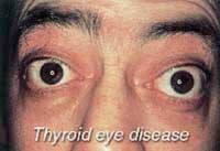
Orbital diseases involve the tissues lying in the bony socket. Generally, the eyeball protrudes from its socket, producing a widening of the eyelids. Sometimes the patient does not blink frequently, developing a staring gaze. This may be the result of an endocrine disorder (thyroid disease), inflammation of the orbit or a tumor. Generally, these lesions require investigations including CT scan and MRI. Treatment varies from case to case and may involve medical treatment, surgery, radiotherapy, chemotherapy or a combination of these.
Know More...
A suspicious lid mass needs excision, examination under microscope and reconstruction of the resultant lid defect. Histopathological examination determines whether the lesion is cancerous or not, and the chances of its recurrence. Reconstruction in the form of suturing, tissue flaps from neighboring areas & other lid, and grafts preserve the lid function.
Know More...
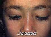
Apart from being cosmetically unacceptable, any irregularity of the lid margin is functionally detrimental to the eye, as lid defects may fail to cover the cornea fully and provide adequate lubrication. An oculoplastic surgeon repairs the injury in a way to make the lid as close to normal as possible.
Know More...
Normally tears from the eye drain to the nose through the lacrimal passage. In case of any blockade in this passage, watering results. The causes can be incomplete development, seen in young children, or infection, which occurs in adult life. Treatment varies from performing a relatively simple procedure like 'probing' the pathway to open it, to more complex surgery of fashioning an alternative pathway to drain the tears to nasal cavity. This procedure is known as dacryocystorhinostomy (DCR).
Know More...
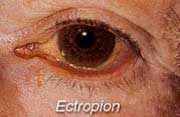
Ectropion is the opposite condition, and the lower lid usually turns away from the eyeball. Ectropion may be due to laxity of the tissue in elderly people or to paralysis of the seventh cranial nerve (the nerve which controls the facial expressions), which causes the weakness of the muscles of the lid. It may also follow cuts, infections, or burns of the lids and face that heal poorly; the resultant scar tissue forms adhesions that cause the lids to turn out. Besides being cosmetically unpleasant, ectropion is accompanied by troublesome tearing and infection. Treatment is surgical rotation of the lid margin and its alignment with the eyeball.
Know More...
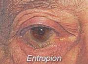
In a condition known as entropion there is inward turning of the eyelids, causing the eyelashes to scratch the cornea and produce irritation. Tearing and secondary infection as well as an unpleasant looking eye cause the patient to seek medical care. Entropion may be the result of spasm or secondary contracture or strictures from burns, injury or trachoma infection. It may involve the upper or lower lids. An adhesive tape applied to the skin of the lid temporarily may straighten the lid and relive the annoying symptoms. Corrective surgery is usually required for a permanent cure.
Know More...
Trichiasis is a condition in which there is misdirection of eyelashes. If the eyelashes turn in toward the eyeball and scratch the cornea, they produce a sensation like a foreign body. This condition may result from trachoma (an eye infection), burns or injuries to the lids. Removal of the offending lashes or corrective plastic surgery on the lid relieves the symptoms.
Know More...
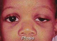
'Drooping' of the eyelid can be present from birth or develop later in old age. It is a cosmetic blemish but if severe, it restricts vision as well. The treatment in majority of cases consists of surgical correction. Surgery involves either strengthening the muscle, which elevates the lid, called LPS resection, or lifting up the lid with the help of a graft. This graft can be taken from the patient's thigh area or can be an artificial sling material. This procedure is known as 'Frontalis Sling'.
When ptosis occurs in adults, it may be the result of a systemic disease, such as myasthenia gravis, which can be treated medically. It can also follow muscle or nerve damage in other parts of the body, or tumors of the lid. When ptosis occurs suddenly in one eye, disease of the brain itself must be considered, and the patient should be seen at once by a neurologist.
Know More...
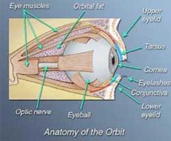
Before some of the common problems are discussed, it is important to understand the anatomy of the structures around the eyeball. The delicate structure of the eyeball is protected, against injury, on the sides and in front by bony walls of the orbit and eyelids respectively.
The orbit is the bony cage or the socket in the skull, which houses the eye. In front of the eye, the eyelids open and close by reflex or voluntary action to distribute tear fluid so as to keep the cornea (the front surface of the eye) moist, to shut out light, and to protect the eyes from foreign bodies and exposure. The outer surface of the lids is a layer of skin continuous with the skin of forehead above and that of the cheeks below. The outer layer of the lids contain muscles that elevate and lower the lids, a firm tissue plate, or tarsus, that maintains their shape, and eyelashes that prevent perspiration or small foreign bodies from entering the eye and damaging the transparent sensitive surface of the cornea. The inner surface of the lids is lined by a mucous membrane called the conjunctiva; this is continuous with the conjunctiva covering the white of the eyeball. The conjunctiva has a rich supply of blood vessels, which accounts for the bloodshot appearance of the eye after irritation. It also contains lubricating glands that permit the lids to move easily and the eye to rotate smoothly. Behind the upper eyelids are present the main tear-producing glands (the lacrimal glands).
The orbit contains, apart from the eyeball, nerves, blood vessels, fat, eye-muscles (to move the eyes freely and harmoniously in both directions), and the optic nerve, which transmits visual sensation from the eye to the brain. The orbit also forms the wall to the adjacent sinuses, which are air spaces in the skull lined by the same kind of membrane as the nose. Canals connecting the eyelids to the sinuses (Lacrimal system) allow secretions and tears to drain through the nose. Some of the problems frequently encountered by an Oculoplastic surgeon include:
Know More...
Any painful blind eye needs removal. The deep 'socket' left behind is not ideal for artificial eye fitting. Therefore, at the time of eye removal, an implant is placed in the orbit, which occupies the space taken by the normal eyeball. This reduces the hollowness of the socket seen with the artificial eyes placed without an implant. In some people, the artificial eye fit changes with passage of time. Socket surgery aims at giving the best possible 'bed' for artificial eye fitting, with or without an orbital implant. The above-mentioned list of disorders is by no means exhaustive. Lack of space prevents description of all conditions seen by an oculoplastic surgeon. Do not hesitate to contact your eye specialist for further information.
Know More...
 Dr. Maneesh Kumar
 Dr. Saman Adil


 Call us
 Email us


