
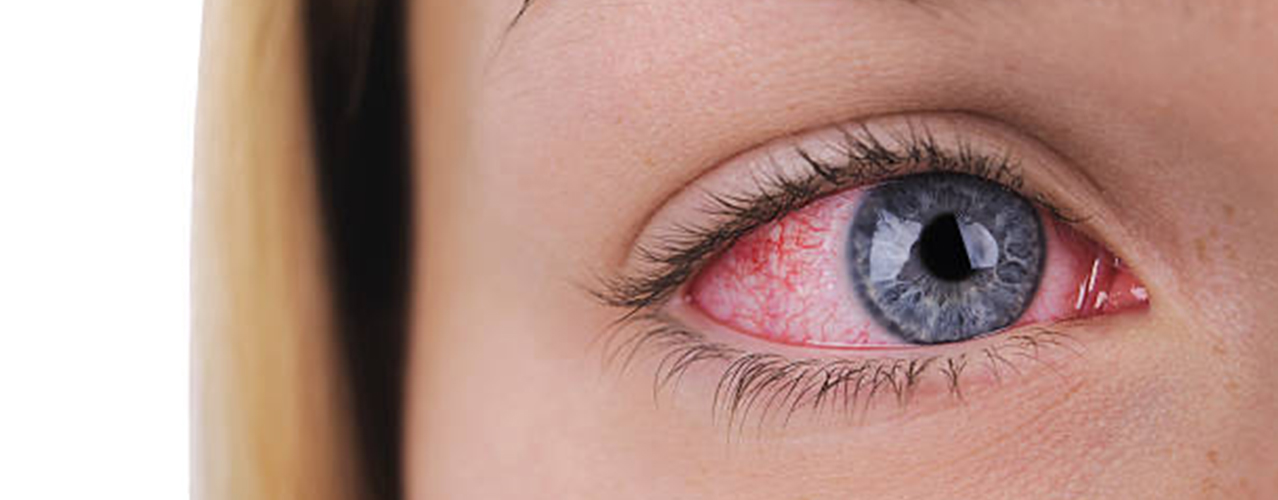
There are two types of vessels in the retina - arteries and veins. A retinal artery occlusion(RAO) is a blockage of one or many arteries/arterioles in the retina. This blockage can result from a clot blocking the vessel lumen or cholesterol lining the vessel wall and eventually blocking it. It can also be considered as a stroke (called by many as an ‘eye stroke’)..
It is an ophthalmic emergency and needs urgent treatment.
Retinal artery occlusion
There are two types of vessels in the retina - arteries and veins. A retinal artery occlusion (RAO) is a blockage of one or many arteries/arterioles in the retina.
This blockage can result from a clot blocking the vessel lumen or cholesterol lining the vessel wall and eventually blocking it. It can also be considered as a stroke.
(called by many as an ‘eye stroke’).
It is an ophthalmic emergency and needs urgent treatment.
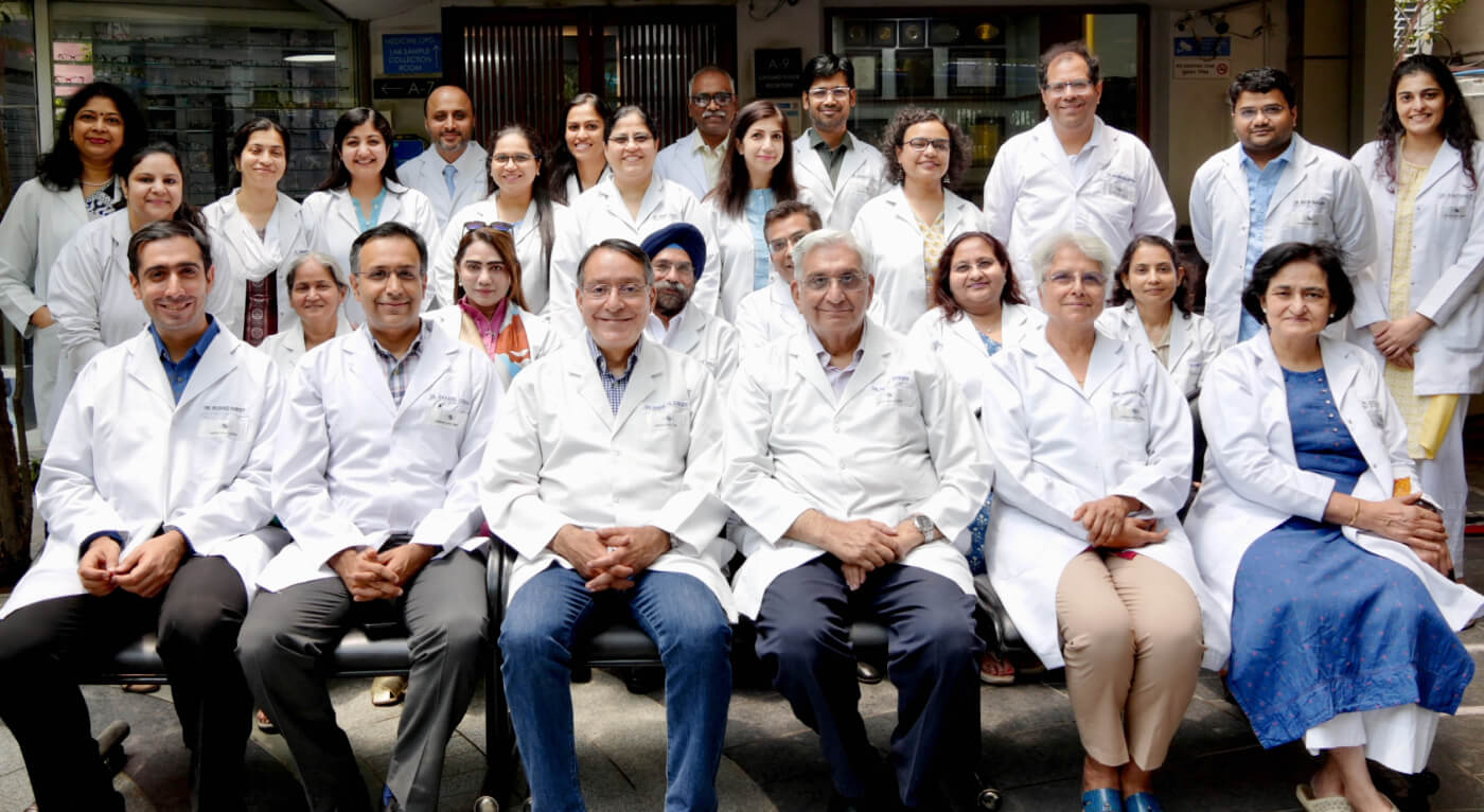
why choose
shroff eye centre
for your treatment?
LEADERS IN EYE CARE SINCE 1914
-
NABH ACCREDITED
-

ISO 9001:2015
Accredited -

ZEE HEALTHCARE
LEADERSHIP AWARD -

Radio City Icons
Award -

AWARDED TRUSTED
EYE CENTRE -

Times Health Survery
Top Eye Clinics in
North India & Delhi 
best eye hospitalS
in north india


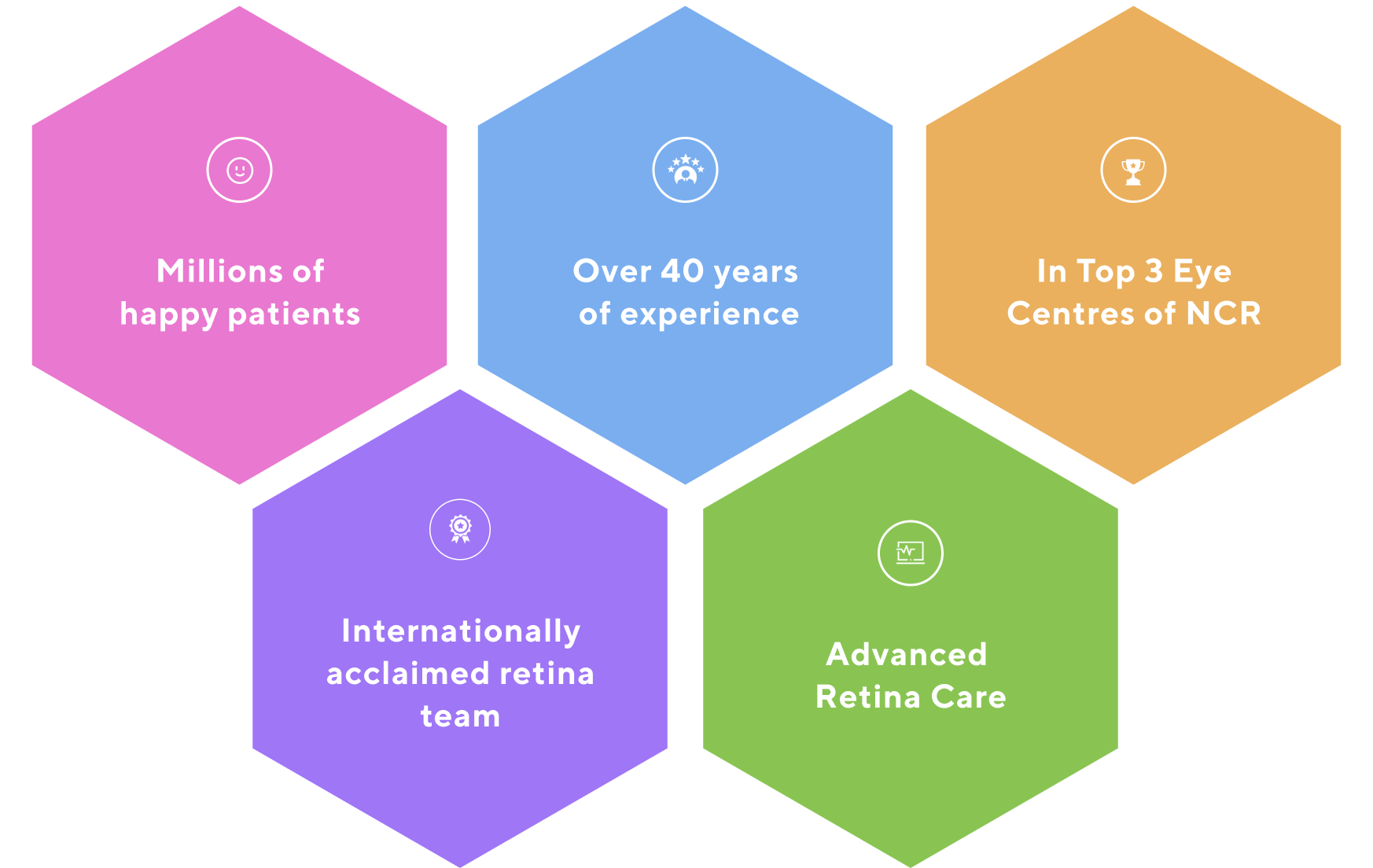

Millions of
happy patients

Over 40 years
of experience

In Top 3 Eye
Centres of NCR

Internationally
acclaimed retina
team

Advanced
Retina Care
TESTIMONIALS
Don’t just take our word. Here’s what patients have to say about us!
I was delighted to visit Shroff’s Eye Hospital and see that it has been refurbished and expanded in a Spacious and beautiful manner. This well equipped and competently staffed Eye Centre has become a facility of excellence which will be a boon to the people of Delhi. I wish the Shroff Eye Centre the people and saving their eye sight.
Shri. K. R. Narayanan, President of India
(1997-2002), Rashtrapati
Bhawan
I am of firm opinion that Dr. Shroff’s institution is the best eye institution. I am fully satisfied with the care and treatment of my eyes. I had heard lot of praise about this institution, I got the same. I wish all the best for this institution.
Shri. Raj Nath Singh,
Defence Minister of India
This is undoubtedly the finest institution for eyes. Not only is every single staff member highly professional and brilliantly competent, everyone is warm, patient understanding and patient friendly. God bless all of you.
Shri. Prannoy Roy
Founder, NDTV India
Successful retina detachment Surgery in Delhi NCR
Rajiv Miglani
Diabetic Retinopathy ke badiya doctor
Dr. Daraius Shroff
happy patients
Successful retina detachment Surgery in Delhi NCR
Rajiv Miglani
Diabetic Retinopathy ke badiya doctor
Dr. Daraius Shroff


BASED ON 5K+ REVIEWS

mukul giri
We had our Retina Detachment surgery. Dr Shroff is the best retina surgeon and the surgery was done with utmost precautions and results were outstanding.

Bhaskar Azad
Excellent services and great care. The whole staff was very professional and took great care of my eye - I had a retina issue which was resolved completely.

Gauri Jain
Gagan Bhatia sir is the Best doctor in East Delhi for Retina checkup. We are satisfied with his treatment..Very polite and well versed.

Google Rating
4.9

5000+
Reviews
Types of
Retinal Artery Occlusion
RAOs can be divided into two types depending on the type of vessel blocked.
-
Blockage of smaller arteries (arterioles) results in a Branch Retinal artery Occlusion (BRAO).
-
Occlusion of the major retinal artery I.e. central retinal artery results in Central Retinal Artery Occlusion (CRAO).


Risk factors of Retinal Artery Occlusion
Systemic risk factors include:
-
High blood pressure
-
Uncontrolled blood sugar
-
Elevated cholesterol
-
Smoking
-
Coronary artery disease
-
Carotid artery stenosis
-
Cardiac arrhythmias
Other risk factors include:
-
Inflammation in the vessel walls
-
Coagulation disorders
-
Leukemias/Lymphomas
what are the Types of Retinal artery Occlusions?
RAOs can be divided into two types depending on the type of vessel blocked.
Blockage of smaller arteries (arterioles) results in a Branch Retinal artery Occlusion (BRAO).
Occlusion of the major retinal artery I.e. central retinal artery results in Central Retinal Artery Occlusion (CRAO).
Central Retinal Artery Occlusion (CRAO)
Branch Retinal artery Occlusion (BRAO)
Risk Factors for Retinal artery Occlusion
Systemic risk factors include:
-
High blood pressure
-
Uncontrolled blood sugar
-
Elevated cholesterol
-
Smoking
-
Coronary artery disease
-
Carotid artery stenosis
-
Cardiac arrhythmias
Other risk factors include:
-
Inflammation in the vessel walls
-
Coagulation disorders
-
Leukemias/Lymphomas
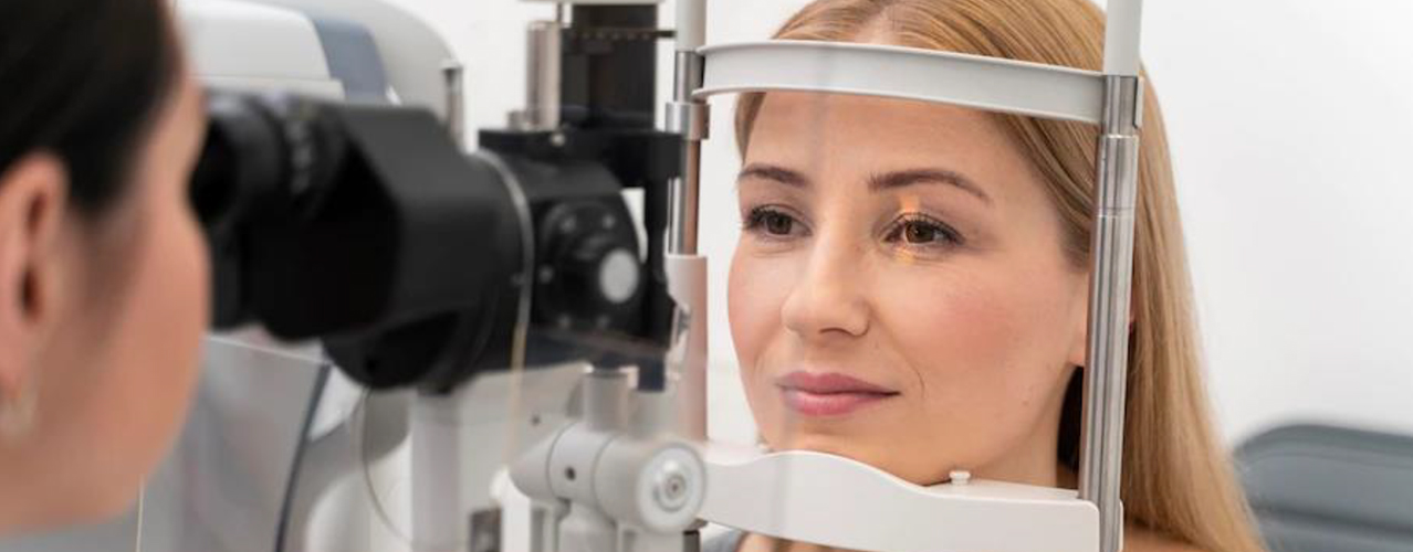
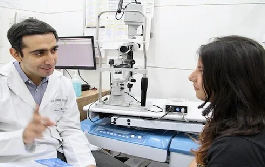
Retinal Artery Occlusion diagnosis

In case someone develops the above symptoms, they should contact an ophthalmologist immediately and avoid any delay in initiating treatment. Diagnosis involves:
-
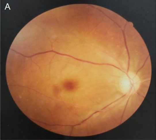
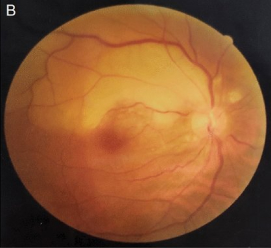
A good and thorough clinical examination including a dilated retina exam. A classical finding in CRAO is a “Cherry red spot” at the macula.
-
Fundus fluoroscein angiography(FFA) to assess the circulation of the retina to check for signs of blockage and reduced retinal perfusion.
-
Optical Coherence Tomography (OCT) shows swelling and thickening of the retinal layers in an acute occlusion.
-
Fundus Photograph: A picture of the retina may be taken to document the condition of the retina and it allows us to monitor the retina as well.
Other investigations including a full systemic workup and a cardiologist and/ or neurologist are also suggested. Tests like ECG, Echocardiography, blood investigations, carotid doppler (ultrasound of the neck vessels) to rule out systemic risk factors.
Draining fluid from the anterior chamber of the eye to induce hypotony - in order to dislodge any clot if present
-
If initiated early (within a few hours), there is a chance that the retinal perfusion may be restored to some extent. However the prognosis remains guarded as most patients do not reach a doctor early.
Treatment modalities include :
-
Lowering the eye pressure using IOP lowering drugs
-
Digital ocular massage
-
Draining fluid from the anterior chamber of the eye to induce hypotony - in order to dislodge any clot if present
-
Further treatment including vitrectomy has been tried in a few patients under guarded prognosis.
-
Hyperbaric oxygen therapy is being tried as a newer modality to help treat recent CRAO.
Your cardiologist or neurologist may start blood thinner and cholesterol lowering agents as well.
RAO is unpredicatable and no one can prevent its occurrence.
However, a good metabolic control of blood pressure, blood sugar and cholesterol goes a long way in preventing such serious conditions.
Avoidance of smoking is a major factor which is known to reduce the risk of RAO.
A healthy lifestyle, regular exercise and frequent follow ups are important ways by which one can maintain a healthy eyesight and long life.
In case someone develops the above symptoms, they should contact an ophthalmologist immediately and avoid any delay in initiating treatment. Diagnosis involves:
Comprehensive dilated retina exam
A good and thorough clinical examination including a dilated retina exam. A classical finding in CRAO is a “Cherry red spot” at the macula.
Fundus fluoroscein angiography (FFA)
to assess the circulation of the retina to check for signs of blockage and reduced retinal perfusion.
Optical Coherence Tomography (OCT)
shows swelling and thickening of the retinal layers in an acute occlusion.
Fundus Photograph
A picture of the retina may be taken to document the condition of the retina and it allows us to monitor the retina as well.
Other investigations including a full systemic workup and a cardiologist and/ or neurologist are also suggested. Tests like ECG, Echocardiography, blood investigations, carotid doppler (ultrasound of the neck vessels) to rule out systemic risk factors.
Treatment OF Retinal artery Occlusion
RAO is an emergency and needs immediate intervention.
If initiated early (within a few hours), there is a chance that the retinal perfusion may be restored to some extent.
However the prognosis remains guarded as most patients do not reach a doctor early.
Treatment modalities include :
-
Lowering the eye pressure using IOP lowering drugs
-
Digital ocular massage
-
Draining fluid from the anterior chamber of the eye to induce hypotony - in order to dislodge any clot if present
-
Further treatment including vitrectomy has been tried in a few patients under guarded prognosis.
-
Hyperbaric oxygen therapy is being tried as a newer modality to help treat recent CRAO.
Your cardiologist or neurologist may start blood thinner and cholesterol lowering agents as well.
Prevention of Retinal Artery Occlusion
RAO is unpredicatable and no one can prevent its occurrence.
-

However, a good metabolic control of blood pressure, blood sugar and cholesterol goes a long way in preventing such serious conditions.
-

Avoidance of smoking is a major factor which is known to reduce the risk of RAO.
-

A healthy lifestyle, regular exercise and frequent follow ups are important ways by which one can maintain a healthy eyesight and long life.
Frequently Asked Questions
Why does retinal vein occlusion affect eyesight?
The nerve cells of the retina convert light signals into electrical signals and transfer this to the brain to interpret.
These nerve cells need nutrients and oxygen to function- this is brought by the retinal artery. The metabolites and carbon dioxide are carried away by the retinal vein.
Any blockage due to pressure from atherosclerosis (deposition of cholesterol and plaques inside vessels) in retinal artery or blood clots can cause retinal vein occlusion.
When there is a blockage, the blood cells and the cells of the retinal vein walls release certain chemicals. These chemicals produce edema and promote new blood vessels formation which affects eyesight by causing:
-
Macular Edema- The edema in the macula (the central most part of the retina responsible for fine vision)
-
Neovascularisation- proliferation of new, abnormal blood vessels on the retina. These new vessels are leaky and leak fluid and blood into the vitreous. This can lead to floaters, vitreous haemorrhage and even retinal detachment.
-
Neovascular glaucoma- rise in eye pressure due to new growth of blood vessels near the angle of the eye.
Blindness- If untreated, CRVO can cause irreversible blindness
image gallery


Other services @ SEC

LASIK &
Refractive
Surgery

Dry Eye

Cataract

Glaucoma

Pediatric
Ophthalmology

Squint
Orthoptics

Uvea

Cornea
follow @shroffeyecentre
on instagram
News & events

Do's and Don'ts of wearing and removing specsDr. Rushad Shroff is a Cornea, Refractive and Cataract Surgeon at Shroff Eye Centre in...


 Call Now
 Book an
 Chat
Chat  Our
Our