


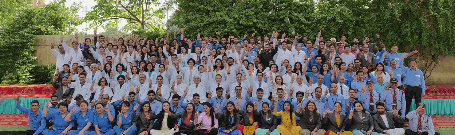

Leaders in Eye care since 1914
-
100 years &
4 generations in Eye Care -
1,00,000+
patients per year -
10,000+
surgeries per year -
4 Centres
-
12 Super specialties
-
36 eye specialists
Shroff Eye Centre is a premier, multi speciality, eye hospital in North India. We offer world-class modern diagnostic, therapeutic, and rehabilitative services of the highest quality.


Shroff Eye Care was established in 1914 in Delhi.
With 4 eye clinics in Delhi, Gurgaon & Ghaziabad, Shroff Eye Centre is amongst the oldest and most reputed ophthalmology practices in the country.
A tertiary eye centre, over one lakh patients visit Shroff Eye centre and over ten thousand eye surgeries are performed every year.
A firm commitment to quality and ethics with the best patient care possible, is at the heart of all services provided at Shroff Eye Centre.
Our services


I've been a patient here for 20 years, since ai first needed glasses. My power finally stabilised last year so I underwent Lasik (Silk) and my experience was fantastic. It was quick, 20 minutes tops, and painless. Dr Rushad was very efficient and knowledgeable.
Neeti Banerji
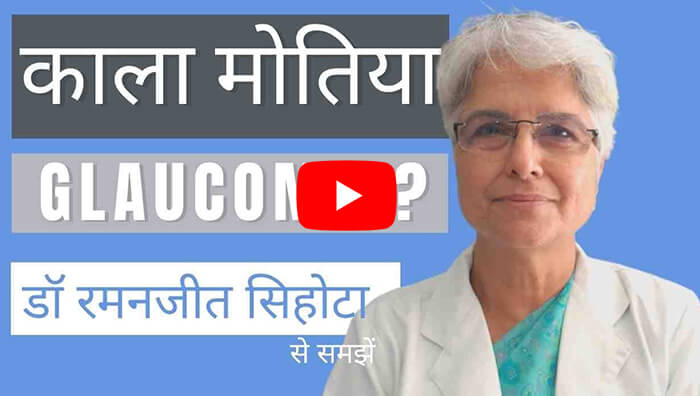
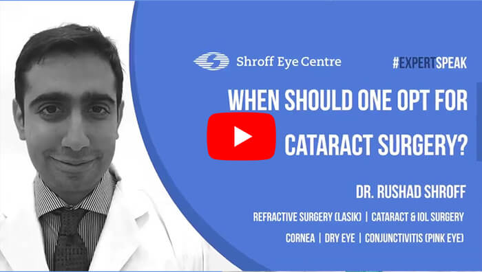
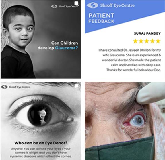
image gallery
@ shroffeyecentre


follow @shroffeyecentre
on instagram
News & events
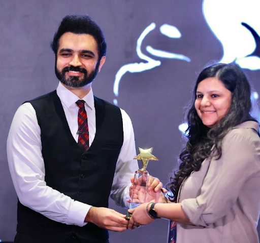
Dr. Apoorva Agrawal Receives Best Film Award for Innovative Dry Eye Diagnosis Presentation!Celebrating the remarkable achievement of our own Dr. Apoorva Agrawal, who clinched the Best Film...
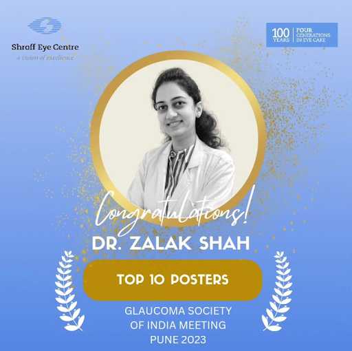
Dr. Zalak Shah Recognized Among Top 6 in India: Selected for Oral Presentation at Glaucoma Society of India Meeting!We’re delighted to share the outstanding achievement of our Glaucoma Fellow, Dr. Zalak Shah, whose...

Congratulations to Dr. Apoorva Agarwal: Winner of the Best Video in Refractive Surgery at IIRSI Conference 2023!We’re thrilled to announce that Dr. Apoorva Agarwal secured the prestigious Best Video in Refractive...


 Call Now
 Book an
 Chat
Chat